Introduction to Canine Cardiac Diseases

by Vickie Halstead, RN, CCRN, CEN, CVNS, LNC
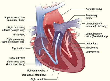
Diagram from The National Heart, Lung, and Blood Institute
www.nhlbi.nih.gov/health/dci/Diseases/pda/pda_heartworks.html
This is the first article in a series I will be writing for you on canine cardiac diseases. This article will give you an overview of cardiac anatomy and physiology, which will help you understand the diseases as they are presented in forthcoming articles. Keep this article handy for review when you read the future articles. My hope is that this information will encourage Bichon breeders to obtain the OFA cardiac certification prior to breeding, and will help improve the health of our beloved Bichons.
To start with the basics, the heart is a muscle that functions as a pump. The pump has 4 chambers that run on electricity, not gasoline. The electrical power is derived from the pacemaker of the heart, which sends the electricity to the heart muscle via a wiring system (nerves, i.e. conduction system). Once stimulated with electricity, the muscle contracts and pumps out its content of blood. The heart muscle has a great burden in that it constantly moves, so if it becomes damaged it can’t be rested like other muscles in your body. Heart failure means the heart muscle is weakened from some type of damage, rendering it unable to empty its contents efficiently.
The 4 chambers of the heart are the right and left atria and the right and left ventricles. These chambers house muscle in their walls, as shown in pink on the above diagram. The chambers communicate via valves that function like trap doors to keep blood flowing in the right direction through the heart. First, deoxygenated blood (blue) is returned from the body via veins, after the tissues and organs have taken the oxygen they need, to the right atrium via the superior vena cava (collects blood from the brain and upper body) and the inferior vena cava (collects blood from the lower body and organs). All of this ‘blue”? blood collects in the right atrium until the atrium is stimulated by the electricity to contract, which opens the tricuspid valve and dumps its contents into the right ventricle. Once the right ventricle is filled with blood it is stimulated by the electrical current to contract, opening the pulmonary valve and sending the ‘blue’ blood to the lungs where the blood is oxygenated and becomes ‘red’. Once the blood is full of oxygen, it’s returned to the left side of the heart (the left atrium) via the pulmonary veins. Once the left atrium is filled with blood and stimulated by the electrical current, it contracts and empties its contents into the left ventricle through the mitral valve. The left ventricle then fills and becomes stimulated to contract by the electrical current, emptying its contents into the aorta through the aortic valve. From the aorta, via arteries, the rich, oxygenated blood (‘red’ ) is pumped to the brain, and to the rest of the body and organs, only to return later to the right side of the heart as ‘blue’ blood. The miracle of this process is that it all occurs within less than a second!
A healthy heart is a very efficient pump. The left side of the heart contains more muscle due to pumping blood to the arteries of the body, which requires high pressure (your blood pressure) to move blood through this high resistance systemic circuit. The right side of the heart does not need to generate high pressure to pump blood only to the lungs, which is a low resistance circuit called the pulmonary circuit. This efficient pump can be modified by diseases that weaken the muscles, damage the valves causing them to leak, alter the conduction system (wiring) which is called an arrhythmia, i.e. an abnormality in the normal rhythm of the electrocardiogram (EKG), or by malformations of the heart and great vessels (aorta and pulmonary artery) that are present at birth. These diseases will be discussed in future articles.
Heart failure means that one or both sides of the heart is not able to pump efficiently due to weakened muscles in the ventricles, which causes blood to back up in the system. To explain it simply, I use a toilet as an analogy, yes a toilet! Imagine a toilet is one side of the heart. If it’s the left side, the tank is the left atrium, the flushing knob is the mitral valve, the bowl is the left ventricle, all the plumbing gadgets in the tank reflect the strength of the muscle, and the sewer pipe below the toilet is the aorta. The goal is to get a good flush, i.e. a good cardiac output that sends plenty of fluid (blood) out the sewer pipe (to the body). If the toilet is the right side of the heart, the tank would be the right atrium, the knob the tricuspid valve, and the bowl the right ventricle, and the sewer pipe the pulmonary artery.
Diseases of the heart can be demonstrated by the affect on the toilet. A good flush could be impaired by:
1. The gadgets are not working together properly (weak muscles)
2. There is too much water in the tank (left atrium)-fluid overload
3. The flushing knob is faulty (mitral valve)
4. There is too much water in the bowl (left ventricle)-fluid overload
5. The sewer pipe is occluded by buildup of excrements (atherosclerosisd)-build up of
cholesterol and plaque
If the toilet has trouble flushing all of its water from the tank or out of the sewer pipe, it backs up out of the toilet bowl onto the floor. In the left side of the heart blood would back up into the lungs causing breathing problems. In the right side of the heart blood would back up into the venous system causing enlarged neck veins and liver, plus edema (swelling due to fluid retention) in the extremities. Chronic heart failure will cause a combination of the above symptoms since both sides of the heart are involved.
There are basically 3 categories of heart diseases in dogs:
1. Congenital (present at birth and genetically transmitted) heart diseases, such as patent ductus arteriosis (PDA)
2. Hereditary diseases that may appear later than birth, such as valve diseases, dilated cardiomyopathy (disese of the cardiac muscle), and sub aortic stenosis (a problem below the aortic valve)
3. Acquired diseases such as coronary artery disease and chronic valve diseases, which also may be hereditary
These diseases will be discussed in future articles, including the prevalence in Bichons. The Health Committee always encourages you to submit information on any Bichons that have cardiac diseases for our data collection, which is of course confidential.
References:
1. Diseases of the Heart by Charles K. Friedberg
2. ‘Matters of the Heart’ by Mara Bovsun. AKC Gazette, October, 2005
3. ‘Facts on Canine Cardiac Health’ by Kevin Schargen. AKC Gazette, March, 2005
4. OFA web site: www.offa.org/cardiac_about.html